back
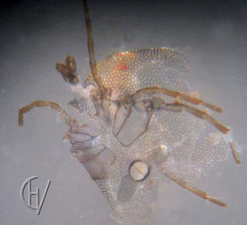
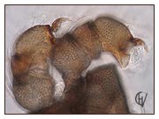
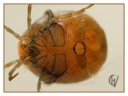
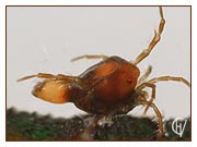
Above is a photo of the dissected skeleton of the water mite described on this and previous pages. The eye spot is still visible. The coaxal plates have a dark edge and their surface appears to be smooth, but they also have a fine-meshed armor, as can be seen in the photo below. Conspicious is the the smooth, white surface of the gonopore membrane, surrounded by a dark edge. The palps have been pulled loose from the skeleton. Note: as a layman I use the word 'armor' and 'skeleton', with that I mean wat is described as the "...completely sclerotized, perforated by large pores" part of the idiosoma (Gerecke, 2016).
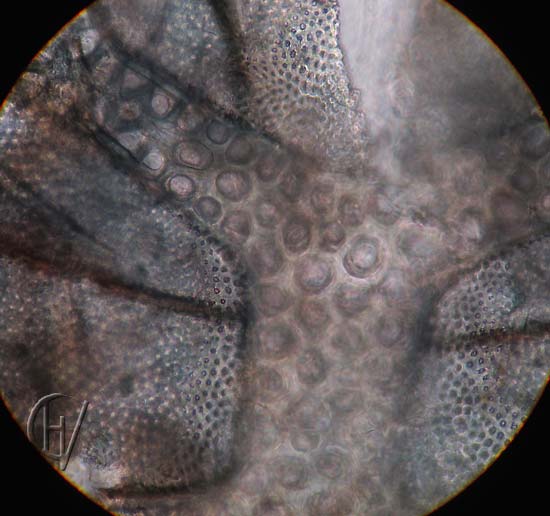
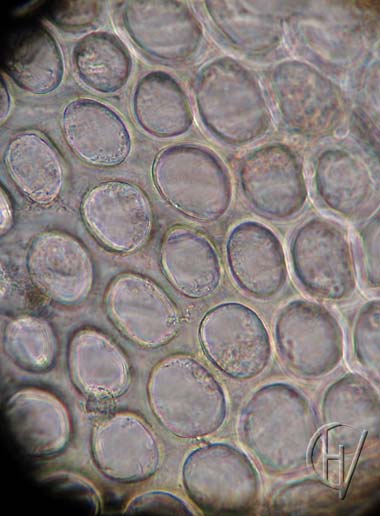
Photo bottom left: the middle section with the epimeres magnified. It is clearly visible that the armoring of the coxal plates is finer and denser than the armoring on the body, which is shown somewhat magnified in the photo bottom right.
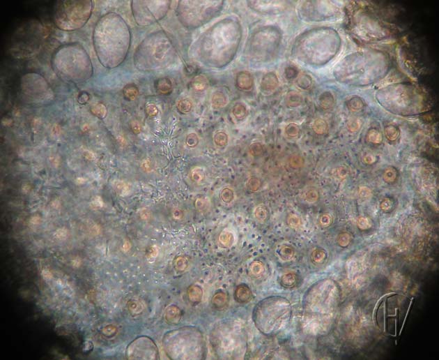
In the photo below one of the cup plates. There seem to be pores between the cups. The armor of the body itself seems to run under the plate, but that is not clearly visible.
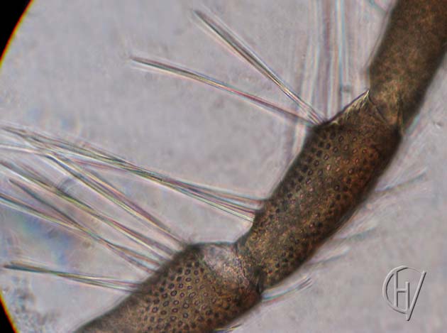
In the photo below part of a leg. The legs also have armor, with small meshes. The leg carries long bristles.