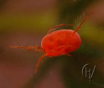
A red watermite (Hydrodroma sp.)

watermite, underside (Piona sp.)
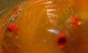
GENITAL Adult watermites bear structures around the genital pore that are
specific for the species. Sometimes these are two movable flaps. On top of, or below
these flaps there are acetabula, which often look like suckerdisclike structures.
Some species have three on either side, other species many more. Below here two
examples:
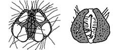
The acetabula were thought to be real suckerdiscs to connect the mating individuals, but that's not true, probably they have an osmoregulating function (Wesenberg Lund, 1939, Davids, 1979). The structure of the genital is an important item for the identification of the species.
The acetabula were thought to be real suckerdiscs to connect the mating individuals, but that's not true, probably they have an osmoregulating function (Wesenberg Lund, 1939, Davids, 1979). The structure of the genital is an important item for the identification of the species.
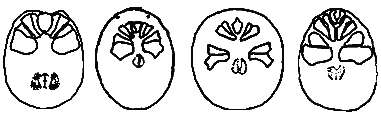
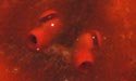
COXAE Chitineous plates on the underside, being the hips (coxa) of the
legs that are fixated to the body. Many species have the coxae fused with the ventral
shield. The shape and position of the coxae are characteristic and differ between
species. Below a few examples, from left to right: Hygrobates, Piona, Hydrodromas and
Arrenurus.
EYES Watermites have two pairs of eyes, on many species these are lying so close
together that there seems to be only one pair. Some species have the eyes on or in
chitineous structures.
four eyes:
Limnesia sp.
Limnesia sp.
two eyes:

Arrenurus sp.
Arrenurus sp.
chitinstructure:
Eylais sp.
Eylais sp.
Is the red colour really a warning?
Proctor H. & Garga N. (2004) found many red watermites in temporary waters without any fish, but in waters were fish lived the not red watermites were dominant. They suggest ”...that the main role of red and orange carotenoid pigments may be to act as photoprotectants, and hypothesize that redness originated in the terrestrial ancestors of water mites.” They further suggest that because red watermites are more conspicuous, they developed the distastefulness. - so this is the opposite of the original idea!
Proctor H. & Garga N. (2004) found many red watermites in temporary waters without any fish, but in waters were fish lived the not red watermites were dominant. They suggest ”...that the main role of red and orange carotenoid pigments may be to act as photoprotectants, and hypothesize that redness originated in the terrestrial ancestors of water mites.” They further suggest that because red watermites are more conspicuous, they developed the distastefulness. - so this is the opposite of the original idea!
Watermites have amazing properties. There are many different species, which are difficult to identify. They are closely related to terrestrial species, and the changes that were needed to live under water are not profound.
The relatively small frontal part, indicated with a yellow colour on the drawing at left, was sometimes called the capitulum (little head), but now mostly
the gnathosoma, (gnatho-: mouthpart, -soma: body). The gnathosoma contains the esophagus and is sometimes stretched, and so forms a snout (rostrum).
The chelicerae (jaws) are the first pair of appendages, but at most species they are are hidden in the gnathosoma, from which they can be protruded. They are build like little spears, not as fangs like the chelicerae of the spiders, but serve to rip through the skin of their prey. Through the surrounding mouthparts, protein solving saliva is then brought in the wound, after which they suck up the liquified tissue. Some species also use the chelicerae to drill holes in waterplants to anchor themselves, Hydrachna species use these holes to inject their eggs.
The palps (as a mites pedipalps are called for short) are the second pair of appendages (marked green on the drawing at left). They are attached to the gnathosoma and on some species might be mistaken for a front pair of legs, or a sort of antennae (ped- foot, palpus feel,touch gently). But they serve several other functions. The palps differ from species to species: long or short, with or without small scissors or pincers. The mites use them to grab and hold their prey, or sometimes as an extra pair of legs to climb or hang from a waterplant. For the identification of a watermite species they are an indispensable help.
The idiosoma is the mites real body (idio-: own, proper) and contains several organs, more on that later. It is not segmented like that of insects and spiders, but shaped like a sack or a globular lump. Hence the name Trombidiformes, (the "lump shaped") for the order in which watermites are placed. A few species have a rather formless body, so weak that these mites will suffer a lethal collapse when brought outside the water, others are more sturdy and some species are really hardened, by an armoured skin. Attached to the lower (ventral) side of the idiosoma are the coxae (hip plates, in red on the sketch at left). The coxae were also called epimeral plates or epimerites for short, so you may find that expression on some of my web pages. When describing a watermite, the pairs of coxae are numbered from 1 to 4, front to back. Coxae 3 and 4 are fused on many species, as are 1 and 2, so then there are four separate fields on the ventral side of the idiosoma, as on the watermite picture at left.
The eight legs, which are the third to sixth pair of appendages, are attached lateral to the coxae, (or may we say the coxae of the legs are fixated to the body?). There are eight legs, many species have fringes or tufts of long hairs on all or some of the legs, which allow them to swim. Most legs have two claws at the end. The males of some species have special leg parts with a function in mating.
Feeding, digestive track. The prey animals are caught with the legs and palps, and after piercing the skin with the chelicerae, liquified protein is sucked up into the stomach (also called mid gut) which has a number of blind ending sacks. This stomach is not connected to a real end gut, but the meal is absorbed into the body. Because their are no insoluble leftovers, a normal anus is not necessary. However, the relative large excretory organ that is lying over and after the stomach, does have an opening to the outside, through which now and then translucent globules are discharged. This opening is situated on the sternal part of the underside and is called the excretory pore, anal pore or simple: anus. The globules are the waste products of the body that are passed to the membrane of the excretory organ. When filled the organ shimmers through the skin as a Y-form, in some species it has fine branches which give the mite an attractive colour pattern.
Breathing. There are no lungs, but tracheae, widely branched, very fine tubes, comparable to those of insects. The two main tubes end in stigma's (little openings) on top of the mouthparts, covered by the skin. Through the skin oxygen from the water is taken in. Watermites never come up to breathe, yet the tracheae are filled with air.
The genital is positioned at the underside of the body (see left).
REFERENCES
Proctor, H.C., Garga, N. (2004). Red, distasteful water mites: did fish make them that
way? Exp. & appl. acarology 2004 34 (1-2) 127-47
Excerpt retrieved from http://www.ncbi.nlm.nih.gov/pubmed/15597605
INTRODUCTION ANATOMY LIFE CYCLE SPECIES TAXONOMY

back to: SPRINGTAILS, SPIDERS, MITES, CRUSTACEANS
Page track: INDEX » mites a.o. » watermites » watermites, anatomy
COPYRIGHT:
All pictures on this site were made by Gerard Visser (Aadorp,
Netherlands), unless stated otherwise. All rights remain with him. These pictures may not
be used for purposes any other than private viewing or printing. Do NOT hardlink to these
pictures or place them on other websites without the author's approval. Should you
need them for purposes which include third parties, you must ask the author permission by
e-mail. People, who want to use this pictures for exhibitions or
publications or educative material are much encouraged to do so, after approval as
mentioned and giving the normal credits.
© G.H. Visser 09-02-2010
rev. 28-04-2022
Deze pagina in het Nederlands
https://www.microcosmos.nl